Table of Contents
Introduction
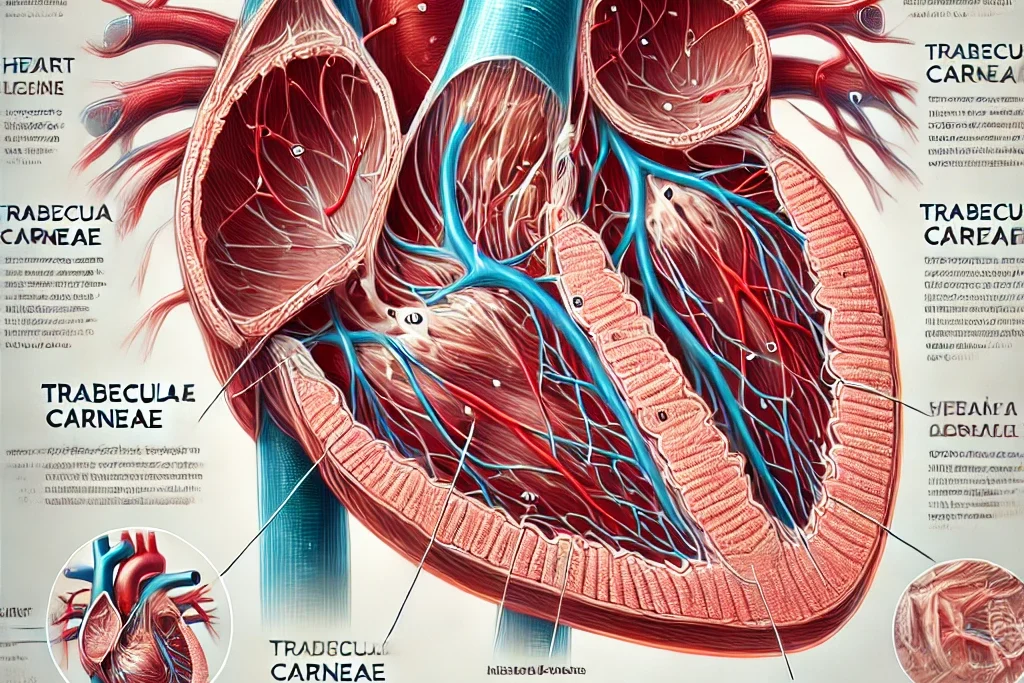
The trabeculae carnea are thread-like muscular ridges located on the inner walls of the heart ventricles. Despite their relatively obscure nature, they play a crucial role in the mechanical and metabolic functions of the heart. This article explores the structure, function, and clinical significance of trabeculae carneae, drawing on various scientific studies to provide a comprehensive overview.

Structure of Trabeculae Carneae
The trabeculae carneae are composed of axially-aligned muscle fibers interspersed with collagenous tissue. According to research by Sands et al. (2011), the collagen content in trabeculae carneae can vary significantly, ranging from 1% to 100% of the cross-sectional area. This variability can influence both the mechanical properties and the metabolic efficiency of the heart muscle.
Imaging Techniques
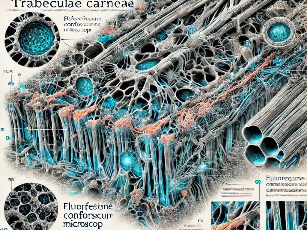
Various imaging techniques, such as fluorescence confocal microscopy and environmental scanning electron microscopy (ESEM), have been employed to study the microstructure of trabeculae carneae. Confocal microscopy provides detailed images of the collagenous structures, while ESEM offers high-resolution images that enhance our understanding of the distribution and density of collagen fibers.

Function of Trabeculae Carneae
Trabeculae carneae are not merely embryological remnants but serve significant mechanical functions. They help in the even distribution of stress across the ventricular walls, particularly in the apex where the myocardium is thinnest and wall stress is highest. Studies have shown that cutting the trabeculae can improve left ventricular diastolic compliance, suggesting their role in maintaining ventricular stiffness and function (Halaney et al., 2017).
Clinical Significance
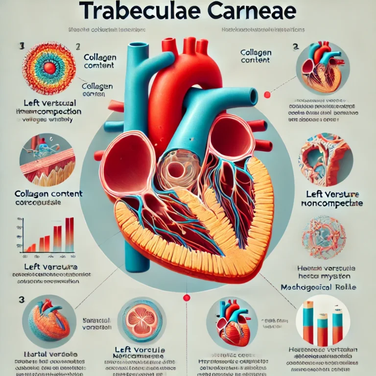
Trabeculae carneae have been implicated in various cardiac conditions. Excessive trabeculation is a hallmark of left ventricular noncompaction, a condition that can lead to heart failure. Furthermore, trabecular hypertrophy is observed in hypertrophic cardiomyopathy and dilated cardiomyopathy. The fibrotic content of trabeculae carneae increases in systolic heart failure, contributing to the overall stiffness and reduced compliance of the ventricles.
Research Insights
- Collagen Content: The collagen fraction in trabeculae carneae varies widely, which can significantly impact the mechanical properties of the heart muscle. Research by Sands et al. (2011) quantified the collagen content using advanced imaging techniques, providing valuable insights into the microstructural composition of trabeculae carneae.
- Mechanical Role: Trabeculae carneae contribute to the mechanical integrity of the heart. The study by Halaney et al. (2017) demonstrated that cutting these structures can enhance diastolic compliance, indicating their role in maintaining ventricular stiffness and function.
- Pathological Conditions: Abnormal trabeculation is associated with various pathological conditions, such as left ventricular noncompaction and hypertrophic cardiomyopathy. These conditions can lead to heart failure if not properly managed.
Conclusion
The trabeculae carneae are vital components of the heart’s internal architecture, playing significant roles in both its mechanical and metabolic functions. Understanding their structure and function can provide deeper insights into cardiac physiology and pathophysiology, aiding in the diagnosis and treatment of various heart conditions. Future research should continue to explore the complexities of these structures to further unravel their contributions to heart health.
Sources
- Sands, G., Goo, S., Gerneke, D., LeGrice, I., & Loiselle, D. (2011). The collagenous microstructure of cardiac ventricular trabeculae carneae. Journal of Structural Biology, 173(1), 110-116. doi:10.1016/j.jsb.2010.06.020
- Halaney, D. L., Sanyal, A., Nafissi, N. A., Escobedo, D., Goros, M., Michalek, J., Acevedo, P. J., Pérez, W., Escobar, G. P., Feldman, M. D., & Han, H. (2017). The Effect of Trabeculae Carneae on Left Ventricular Diastolic Compliance: Improvement in Compliance With Trabecular Cutting. Journal of Biomechanical Engineering, 139(3), 031012. doi:10.1115/1.4035585


Pingback: Understanding the Intertubercular Groove: Anatomy, Pathology, and Clinical Significance - Science is Life
Pingback: Curette and Endocervical Curettage (ECC) - Science is Life
Pingback: Can Dogs Eat Chocolate? - Science is Life
Pingback: What is Leadership? - Science is Life
Pingback: Chordae Tendineae 101: Structures and Functions