Table of Contents
INTRODUCTION
Endocervical curettage (ECC) is a significant procedure in gynecology, widely used to diagnose and manage various uterine conditions. The procedure primarily involves using a surgical instrument called a curette. This guide provides an in-depth overview of ECC, emphasizing the role of the curette, including its purpose, procedural steps, potential risks, and recovery process.
What is Endocervical Curettage (ECC)?
Endocervical curettage (ECC) is a medical procedure involving the removal of tissue from the endocervical canal using a curette. The endocervical canal is the passageway connecting the uterus to the vagina. Tissue collected during ECC is critical for diagnosing conditions such as cervical cancer, polyps, and abnormalities in the uterine lining (Bestel et al., 2019; Damkjaer et al., 2022).
The Role of the Curette in ECC
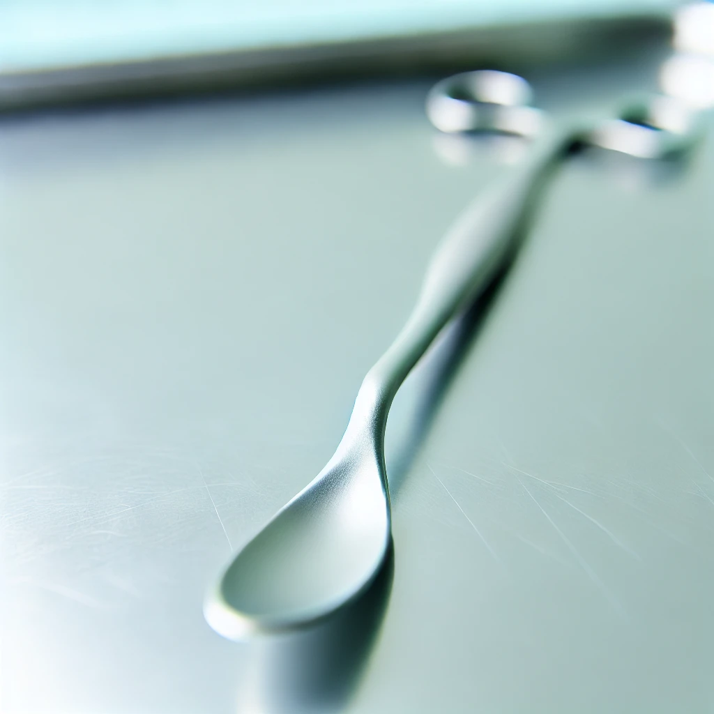
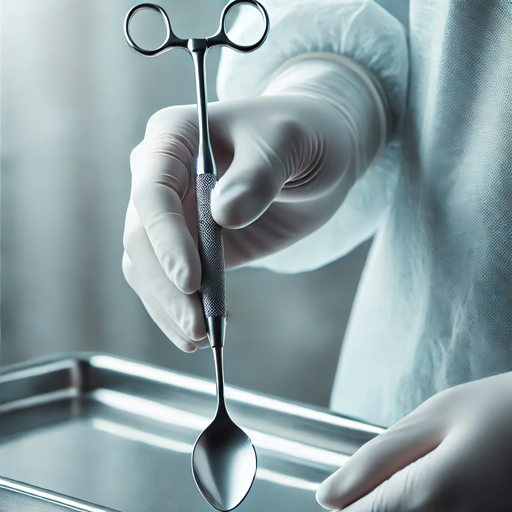
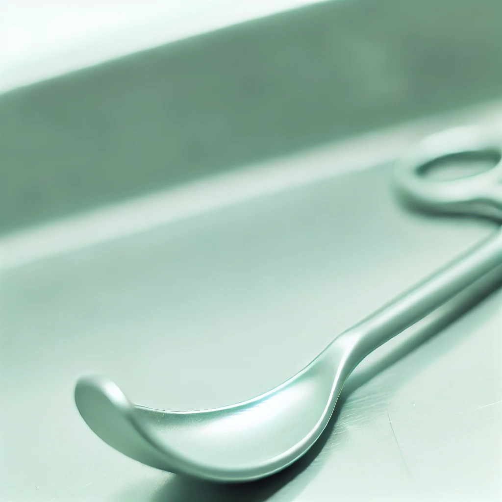
A curette is a spoon-shaped surgical instrument designed for scraping or debriding biological tissue. Its specific shape allows it to effectively and safely remove tissue from the endocervical canal. Curettes come in various sizes and shapes to accommodate different procedural needs and patient anatomies (Goksedef et al., 2013).
When is ECC Performed?
ECC is performed for several reasons, including:
- Abnormal Uterine Bleeding: ECC helps determine the cause of heavy or irregular bleeding that may not be related to menstrual cycles (Damkjaer et al., 2022).
- Cervical Cancer Screening: The procedure is used to detect precancerous or cancerous cells in the endocervical canal (Bestel et al., 2019).
- Polyps or Fibroids: ECC can identify benign growths that might affect fertility or cause discomfort (Goksedef et al., 2013).
- Postmenopausal Bleeding: This procedure investigates the cause of bleeding in women who have gone through menopause (Damkjaer et al., 2022).
Preparation for ECC
Before undergoing ECC, it is essential to review the patient’s medical history. This includes discussing any current medications, allergies, or existing health conditions with the healthcare provider. Proper preparation ensures the procedure’s success and minimizes risks (Perkins et al., 2020).
Read: What is Trabeculae Carnea?
The ECC Procedure: Step-by-Step
- Preparation and Positioning: The patient lies on an examination table, and the healthcare provider explains the procedure. The patient is positioned similarly to a routine pelvic exam (Feltmate & Feldman, 2020).
- Speculum Insertion: The doctor inserts an instrument called a speculum into the vagina to widen it, providing a clear view of the cervix. This step is crucial for accessing the endocervical canal (Feltmate & Feldman, 2020).
- Local Anesthesia: Depending on the patient’s comfort and the procedure’s complexity, local anesthesia may be administered to numb the area, ensuring minimal discomfort (Goksedef et al., 2013).
- Using the Curette: A spoon-shaped instrument called a curette is carefully inserted into the endocervical canal. The healthcare provider gently scrapes the uterine lining to collect tissue samples, which might cause mild cramping similar to menstrual cramps (Bestel et al., 2019).
- Additional Methods: In some cases, a suction device might be used to assist in removing the tissue, especially in procedures like dilation and curettage (D&C), where the cervix is dilated, and the uterine lining is scraped (Damkjaer et al., 2022).

Potential Risks and Complications
While ECC is generally safe, there are some risks involved:
- Infection: As with any invasive procedure, there is a risk of infection. Patients should watch for signs of fever, foul-smelling discharge, or severe pain and contact their healthcare provider if these occur (Page et al., 2021).
- Bleeding: Light bleeding or spotting is common after the procedure, but heavy bleeding should be reported to a doctor (Perkins et al., 2020).
- Uterine Perforation: Rarely, the curette might puncture the uterine wall, leading to more severe complications that require immediate medical attention (Bestel et al., 2019).
- Scar Tissue Formation: Scar tissue, or Asherman’s syndrome, can develop in the uterus, potentially leading to complications in future pregnancies (Perkins et al., 2020).
Recovery and Aftercare
- Immediate Aftercare: After the procedure, patients are usually monitored in a recovery room for a short period. Mild cramping and light bleeding are common, but severe pain should be reported to a healthcare provider (Goksedef et al., 2013).
- Pain Management: Over-the-counter pain relievers, such as ibuprofen, can help manage discomfort. Rest and hydration are crucial during the initial recovery phase (Feltmate & Feldman, 2020).
- Follow-up Appointments: A follow-up appointment is often scheduled to discuss biopsy results and ensure proper healing (Bestel et al., 2019).
The Curette: A Closer Look
The curette is the cornerstone of ECC. Its design is crucial for the effectiveness and safety of the procedure. There are different types of curettes, including:
- Sharp Curettes: These have a sharp edge and are used for more precise scraping. They are particularly useful in removing tough tissue or when a more detailed sample is needed (Maksem, 2006).
- Blunt Curettes: These are less invasive and are used when a gentle scraping is sufficient. They are often used in patients with more sensitive tissues or when minimal tissue removal is required (Klam et al., 2000).
- Suction Curettes: These combine the scraping action with suction to remove tissue more effectively. They are often used in D&C procedures (Damkjaer et al., 2022).
The Importance of Curette Design
The design of the curette is critical for several reasons:
- Effectiveness: The shape and sharpness of the curette ensure that enough tissue is collected for accurate diagnosis (Maksem, 2006).
- Safety: A well-designed curette minimizes the risk of damaging the uterine wall or other surrounding tissues (Gibson et al., 2001).
- Comfort: Modern curettes are designed to minimize discomfort, making the procedure more tolerable for patients (Klam et al., 2000).
Read: Understanding the Intertubercular Groove
Variations in Curette Types
Understanding the different types of curettes and their specific uses can provide deeper insights into their application in ECC:
- Kevorkian Curette: This type of curette is often used for more aggressive scraping and is beneficial in cases where dense tissue needs to be sampled. It has a rigid, sharp edge that can effectively collect sufficient tissue samples (Martin et al., 1995).
- Sims Curette: A versatile curette used for both gentle and more rigorous scraping. It can be employed in various procedures beyond ECC, including biopsies and D&C (Weitzman et al., 1988).
Historical Context and Evolution of Curettes
The development and refinement of curettes have significantly impacted gynecological procedures. Early curettes were rudimentary and often caused more discomfort and higher risk. Over time, advancements in medical technology and a better understanding of gynecological anatomy have led to the development of more sophisticated and safer curettes (Maksem, 2006).

ECC in Clinical Practice
In clinical practice, ECC is often combined with other diagnostic procedures to provide a comprehensive understanding of a patient’s condition. For example, ECC may be performed alongside colposcopy to investigate abnormal Pap smear results more thoroughly (Feltmate & Feldman, 2020).
Patient Perspectives and Experiences
Understanding patient perspectives on ECC can help healthcare providers improve the procedure’s delivery and patient comfort. Many patients report anxiety and discomfort related to the procedure, highlighting the importance of patient education and reassurance (Undurraga et al., 2017). Ensuring patients understand the purpose of the curette and what to expect during and after the procedure can significantly enhance their experience.
Advances in Curette Design and ECC Techniques
Recent advances in curette design and ECC techniques have focused on improving patient comfort and diagnostic accuracy. Innovations include the development of ergonomic handles for better control, sharper edges for precise tissue removal, and materials that reduce discomfort and risk of infection (Klam et al., 2000).
ECC and Future Research Directions
Ongoing research in gynecology continues to explore new materials and designs for curettes to further minimize risks and enhance diagnostic capabilities. Studies are also investigating the integration of imaging technologies with ECC to provide real-time visual guidance during the procedure (Damkjaer et al., 2022).
Conclusion
Endocervical curettage is an indispensable procedure in women’s health, providing critical insights into uterine conditions. The curette’s role is central to the effectiveness and safety of ECC. Understanding the procedure, its benefits, and the recovery process can help patients prepare and recover more effectively. If you’re experiencing symptoms that may require ECC, consult with a healthcare provider to explore this diagnostic option.
References
- Bestel, M., et al. (2019). Agreement between endocervical brush and endocervical curettage in the diagnosis of cervical cancer. Eastern Journal of Medicine, 24(1), 86–90.
- Damkjaer, M., et al. (2022). Endocervical sampling in women with suspected cervical neoplasia: a systematic review and meta-analysis of diagnostic test accuracy studies. American Journal of Obstetrics and Gynecology, 227(6), 839–848.e4.
- Goksedef, B.P., et al. (2013). Diagnostic accuracy of two endocervical sampling methods: randomized controlled trial. Archives of Gynecology and Obstetrics, 287(1), 117–122.
- Page, M.J., et al. (2021). The PRISMA 2020 statement: an updated guideline for reporting systematic reviews. BMJ (Clinical Research ed.), 372, n71.
- Perkins, R.B., et al. (2020). 2019 ASCCP risk-based management consensus guidelines for abnormal cervical cancer screening tests and cancer precursors. Journal of Lower Genital Tract Disease, 24(2), 102–131.
- Feltmate, C. M., Feldman, S. (2020). Colposcopy. Retrieved from https://www.uptodate.com/contents/colposcopy?search=endocervical%20curettage§ionRank=1&usage_type=default&anchor=H11&source=machineLearning&selectedTitle=1~25&display_rank=1#H11
- Maksem, J.A. (2006). Endocervical curetting vs. endocervical brushing as case finding methods. Diagnostic Cytopathology, 34(5), 313–316.
- Klam, S., et al. (2000). Comparison of endocervical curettage and endocervical brushing. Obstetrics and Gynecology, 96(1), 90–94.
- Gibson, C.A., et al. (2001). Endocervical sampling: a comparison of endocervical brush, endocervical curette, and combined brush with curette techniques. Journal of Lower Genital Tract Disease, 5(1), 1–6.
- Martin, D., et al. (1995). Comparison of the endocervical brush and the endocervical curettage for the evaluation of the endocervical canal. Puerto Rico Health Sciences Journal, 14(3), 195–197.
- Weitzman, G.A., et al. (1988). Endocervical brush cytology. An alternative to endocervical curettage? The Journal of Reproductive Medicine, 33(8), 677–683.
- Undurraga, M., et al. (2017). User perception of endocervical sampling: A randomized comparison of endocervical evaluation with the curette vs cytobrush. PLoS One, 12(11), e0186812.


Pingback: What is Cystolitholapaxy? - Science is Life
Pingback: Chordae Tendineae 101: Structures and Functions